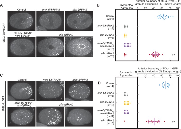
FIGURE 6:
PLK-1 regulates P granule segregation. (A) Spinning disk confocal images of MEG-3::meGFP in embryos of different genotypes at NEBD. Images were taken at the cell midplane. Scale bar = 10 μm. (B, D) Quantification of P granule distribution at NEBD. For embryos in which P granules are asymmetrically distributed, the positions of the anterior-most granules are plotted. Only granules >0.7 μm in diameter were counted to exclude small granules that are sometimes present in the anterior of wild-type embryos. The distribution of P granules in plk-1(RNAi) embryos was significantly shifted toward the anterior compared with the Control (p < 0.0001; Student’s t test). n = number of embryo analyzed. Error bars indicate SD. (C) Maximum projections of spinning disk confocal images of PGL-1::GFP at NEBD. Images were taken at the cell midplane. Scale bar = 10 μm.

