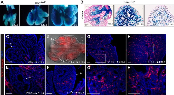
Fig. 1.
Transcription factor 21 (Tcf21) expression in embryonic lung. A: whole mount images of Tcf21LacZ/+ reporter embryonic lungs. B: histologic sections of Tcf21LacZ/+ reporter lungs counterstained with nuclear fast red. Representative images are top left lobe at embryonic day (E)13.5, E15.5, and E18.5; n = 3–5. Scale bars, 50 μm. C–H: Cre-mediated tdTomato expression in embryonic lungs after 1-day labeling. tdTomato expression was visualized using DsRed antibody. Boxed regions in G and H are magnified in G' and H'. D: whole mount fluorescence image of E11.5 lung; lb, lung bud. lb, lung bud; T, trachea. Scale bars, 100 μm.

