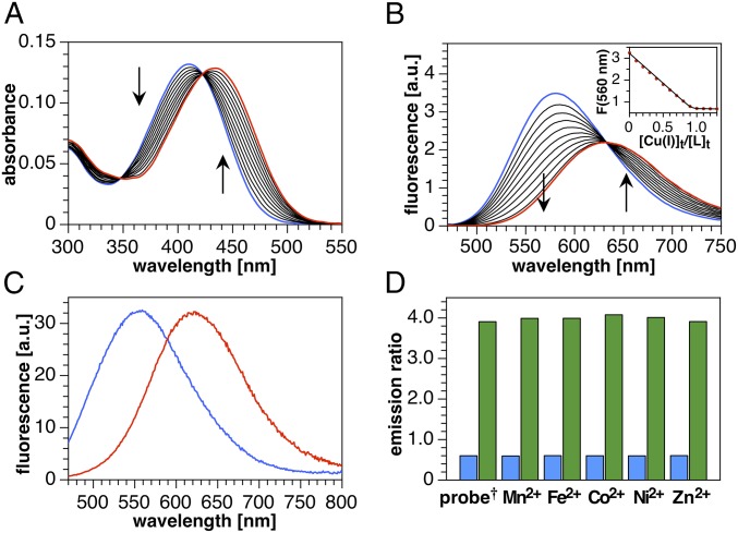
Fig. 3.
Spectral response and selectivity of crisp-17 toward Cu(I) (supplied as [Cu(I)MCL-2]PF6). (A) UV-vis absorption spectra of crisp-17 (5 µM) in 95% MeOH-5% H2O during titration with Cu(I) (0–6.5 µM). (B) Fluorescence emission spectra (λex = 460 nm) under the same conditions. (Inset) Emission intensity at 560 nm vs. molar ratio of Cu(I) to probe. (C) Emission spectra (λex = 450 nm) of crisp-17 (2 µM) equilibrated with liposome suspension (4:1 POPC–POPG, 100 µM total lipids, 10 mM Pipes, pH 7.0, 0.1 M KCl, 25 °C) in the absence (blue trace) and presence (red trace) of 4 µM Cu(I). (D) Ratio of the integrated emission intensities F(590–750 nm)/F(480–580 nm) of crisp-17 (2 µM) in the presence of biologically relevant transition metal ions (10 µM) before (blue bars) and after (green bars) addition of 4 µM Cu(I) (liposome and buffer composition as in C). †Probe response in the absence of a competing metal ion.

