Fig. 4.
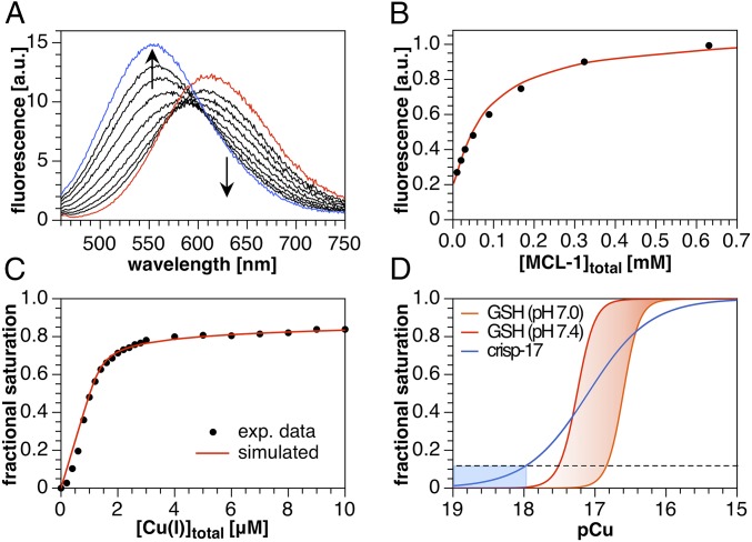
Fluorimetric Cu(I) competition titrations of crisp-17 against MCL-1 and GSH (4:1 DMPC–DMPG, 100 µM total lipids, 10 mM Pipes, pH 7.0, 0.1 M KCl, 25 °C, λex = 450 nm). (A) crisp-17 (1 µM) was equilibrated with 10 µM [Cu(I)MCL-1]PF6 (red trace) at pH 7.0 and subsequently titrated with MCL-1 (10–640 µM, black traces). Full reversibility was confirmed by adding 10 µM PSP-2 (blue trace). Nonlinear least-squares fitting yielded logK of 17.0 ± 0.1. (B) Fit quality evaluated at 530 nm. (C) Apparent fractional saturation of crisp-17, calculated as (I − Ifree)/(Ibound − Ifree), where I represents integrated fluorescence intensity from 500 to 550 nm, upon titration with Cu(I) (as [Cu(I)MCL-2]PF6) in the presence of 4 mM GSH at pH 7.0. Least-squares fitting (red line) yielded logK = 17.1 ± 0.1. (D) Calculated fraction of crisp-17 bound to Cu(I) and of total Cu(I) bound to GSH versus thermodynamically available Cu(I) (pCu = −log[Cu+aq]). The dashed line and blue shading demarcate the Cu(I) availability corresponding to 10% fractional saturation of the probe. The red-shaded area indicates the GSH Cu(I) buffer window within the cytosolic pH range.

