Fig. 6.
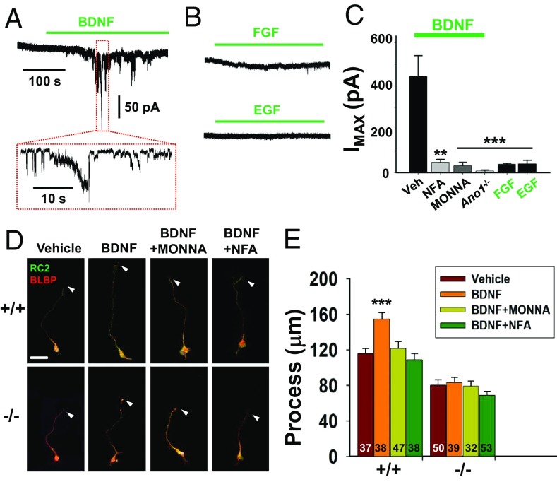
BDNF activates ANO1 in RGCs. (A and B) Representative traces of Cl− current responses of cultured RGCs to BDNF (A) or trophic factors (B). Cells were treated with 100 ng/mL of BDNF, FGF, or EGF. Pipette and bath solutions contained 140 mM CsCl and 140 mM NMDG-Cl, respectively; Ehold = −80 mV. (C) Summary of average current amplitudes of RGCs activated by BDNF, FGF, EGF, or coapplication with 10 μM niflumic acid (NFA) and 30 μM MONNA. The average amplitudes of currents of RGCs isolated from Ano1−/− brains are also shown. **P < 0.01, ***P < 0.001 vs. BDNF (Veh)-evoked currents, one-way ANOVA and Tukey’s post hoc test. (D and E) Representative images (D) and summary of process lengths (E) of RGCs treated with vehicle (Veh), BDNF, BDNF+NFA, or BDNF+MONNA in Ano1+/+ and Ano1−/− RGCs for 3 d. Arrowheads indicate the tips of each process of RGCs. RGCs were stained with BLBP and RC2. (Scale bar: 20 μm.) ***P < 0.001, one-way ANOVA, Newman–Keuls multiple-comparison test.

