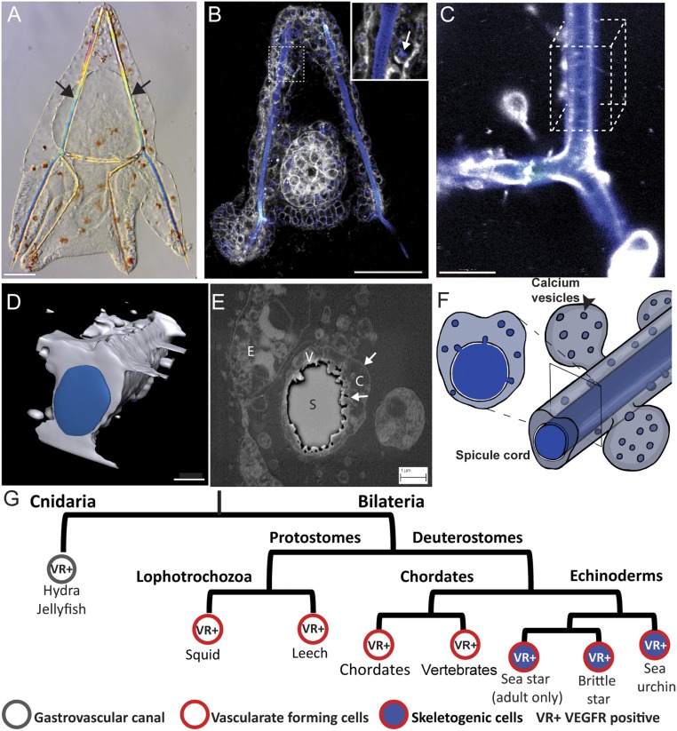
Fig. 1.
Spiculogenesis in the sea urchin embryo and VEGFR expression in tubular organs in metazoan. (A) Sea urchin larva at 3 dpf, showing its two calcite spicules (arrows). (Scale bar, 50 µm.) (B) Live sea urchin embryo at 2 dpf stained with the membrane tracker FM4-64 (gray) and green-calcein that binds to calcium ions (false-colored blue). Enlargement shows the calcium vesicles in the skeletogenic cells (arrow). (Scale bar, 50 µm.) (Enlargement magnification, 400×.) (C) Confocal image of the spicule in live embryo at 3 dpf stained with blue calcein (blue) and FM4-64 (gray). (Scale bar, 10 µm.) (D) Three-dimensional model of the spicule structure based on 50 confocal z-stacks of the cube in C. (Scale bar, 3 µm.) (E) Scanning electron micrograph of a cross-section of the spicule at 2 dpf showing the double membrane cytoplasmic cord that surrounds the spicule and the vesicles inside the cord. C, cytoplasm; E, ectodermal cell; S, spicule; V, vesicles, arrows point to the membranes. (Scale bar, 1 µm.) (F) Schematic model of the spicule and vesicle secretion, showing the calcium vesicles and spicule in blue and cytoplasm in gray. Image courtesy of Yarden Ben-Tabou de-Leon (artist). (G) Partial phylogenetic tree presenting VEGFR expression in cells that generate tubular structures in different phyla throughout the animal kingdom.

