FIGURE 1:
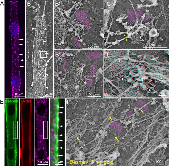
Clathrin-coated plaques anchor desmin IFs. (A) Immunofluorescent staining of CHC (magenta) in differentiated mouse primary myotubes. (B) Survey view of an unroofed primary mouse myotube. (B′, B″) Higher-magnification views corresponding to the boxed regions in B. (C, D) Higher-magnification views of clathrin plaques and associated cytoskeletal structures in unroofed control primary myotubes. Intermediate filaments are indicated with yellow arrowheads and actin filaments are indicated using arrows. For D, use glasses for 3D viewing of anaglyph (left eye = red). (E) Immunofluorescent staining of desmin (green), actin (red), and CHC (magenta) in mouse primary myotubes. (F) Higher-magnification view of clathrin plaques and associated cytoskeletal structures in unroofed control primary myotubes immunolabeled using a primary antibody against desmin and secondary antibodies coupled to 15 nm gold beads. Images are representative of at least four independent experiments. Gold beads are pseudocolored in yellow, and some are indicated with yellow arrowheads. Clathrin-coated structures are highlighted in purple.

