FIGURE 2:
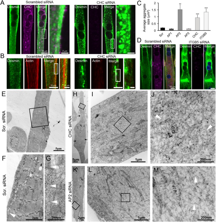
Clathrin-coated plaques are required for desmin IF organization. (A) Immunofluorescent staining of desmin (green) and CHC (magenta) in mouse primary myotubes treated with control or CHC siRNA. Images are representative of at least 10 independent experiments. (B) Desmin (green) and actin staining (red) in mouse primary myotubes treated with control or CHC siRNA. (C) Average desmin aggregate size in myotubes treated with control siRNA or siRNA against CHC, AP1, AP2, AP3, or b5 integrin (ITGB5) (n = 30–50 myotubes). Data presented as mean ± SEM; **, p < 0.01, ***, p < 0.001 using a two-tailed Student’s t test). (D) Immunofluorescent staining of desmin (green) and CHC (magenta) in mouse primary myotubes treated with control or ITGB5 siRNA. (E–L) Thin-section EM of primary myotubes treated with control (E–G), CHC (H–J), or AP2 (K–M) siRNA. I and L are higher-magnification views of IF tangles from H and K, respectively. Images are representative of at least two to four independent experiments. IFs are indicated with arrowheads.

