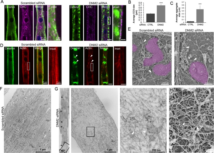
FIGURE 3:
DNM2 is required for desmin and actin organization around clathrin plaques. (A) Immunofluorescent staining of desmin (green) and actin (red) in mouse primary myotubes treated with control or DNM2 siRNA. Images are representative of at least seven independent experiments. (B) Average clathrin-coated structure (CCS) size in myotubes treated with control or siRNA against DNM2 (n = 20 myotubes). (C) Quantification of desmin aggregate fluorescence in myotubes treated with control or DNM2 siRNA (n = 20 myotubes). (D) Desmin (green) and actin staining (red) in mouse primary myotubes treated with control or DNM2 siRNA. (E) High-magnification view of unroofed primary mouse myotubes treated with control or DNM2 siRNA. Actin structures are indicated using arrows. Images are representative of at least 12 independent experiments. (F, G) Thin-section EM of extensively differentiated control (F) or DNM2-depleted (G) myotubes. Images are representative of at least eight independent experiments. Arrowheads denote bone fide IFs. (H) High-magnification view of a desmin aggregate from unroofed primary mouse myotubes treated with DNM2 siRNA. Data are presented as mean ± SEM; ***, p < 0.001, ****, p < 0.0001, using a two-tailed Student’s t test.

