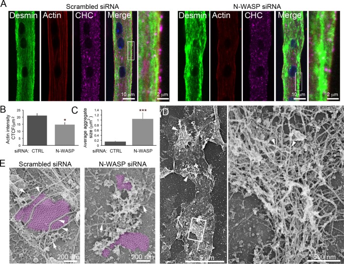
FIGURE 4:
N-WASP is indispensable for desmin and actin organization around clathrin plaques. (A) Immunofluorescent staining of desmin (green), CHC (magenta), and actin (red) in mouse primary myotubes treated with control or N-WASP siRNA. Images are representative of at least five independent experiments. (B) Quantification of cortical actin fluorescence intensity in myotubes treated with control or N-WASP siRNA (n = 20 myotubes). (C) Quantification of desmin aggregate size in myotubes treated with control or N-WASP siRNA (n = 20 myotubes). (D) Survey view of desmin aggregates (arrowheads) from unroofed primary mouse myotubes treated with N-WASP siRNA. (E) High-magnification views of unroofed primary mouse myotubes treated with control or N-WASP siRNA. IFs are indicated with arrowheads, and actin structures are indicated using arrows. Images are representative of at least five independent experiments. Data are presented as mean ± SEM; *, p < 0.05, ***, p < 0.001, using a two-tailed Student’s t test.

