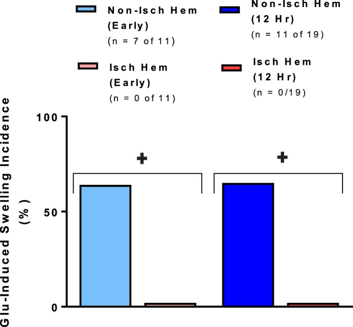
Fig. 3.

Reduced swelling as measured by increased change in light transmittance (ΔLT) induced by bath application of 500 µM glutamate in the predicted ischemic core region of brain slices harvested both immediately and 12 h after middle cerebral artery occlusion. The ischemic hemisphere displays a significantly lower incidence of glutamate-induced tissue swelling that exceeds a 5% threshold Mann-Whitney U-test, (+P < 0.001) than the homotypic contralateral region. Values are means; n = no. of slices. Isch hem, ischemic hemisphere; Non-isch hem, nonischemic hemisphere.
