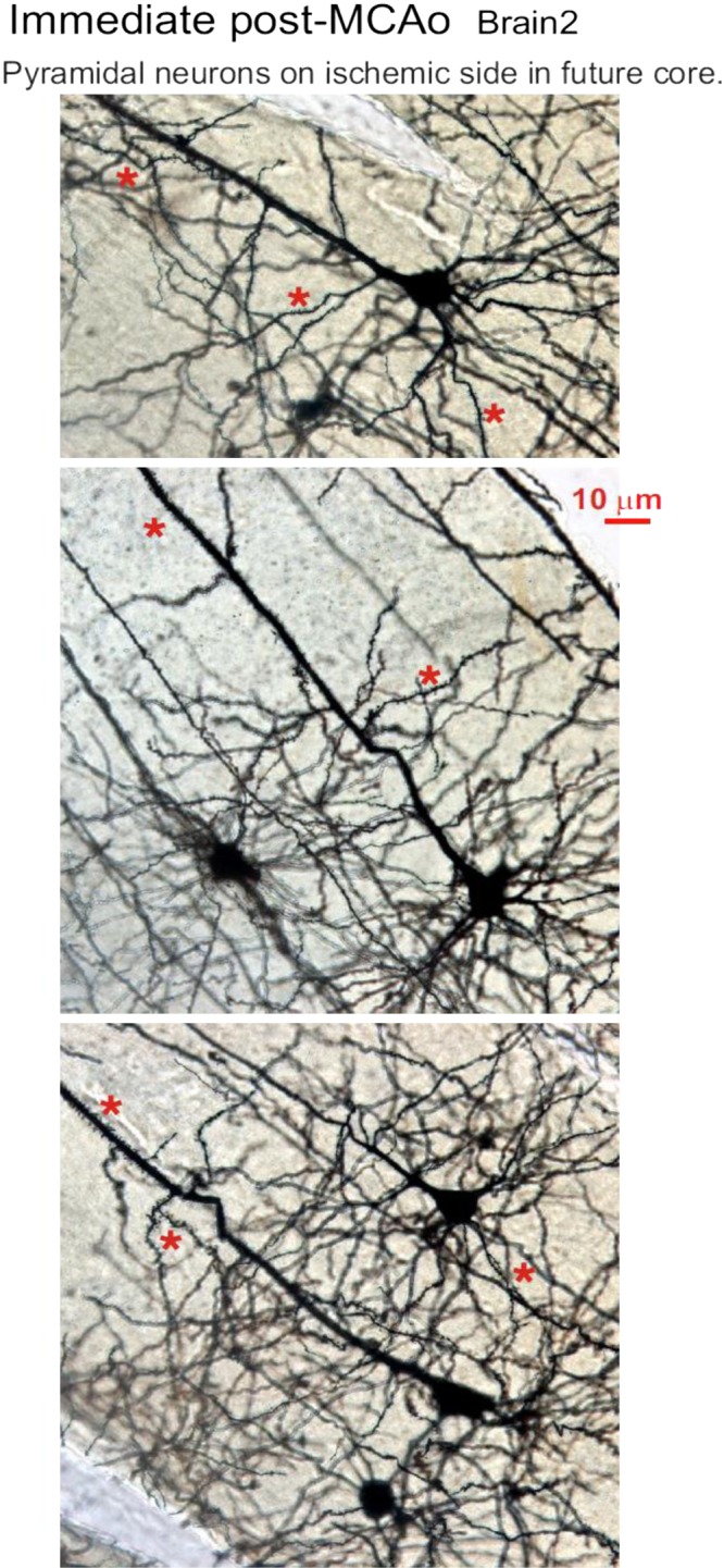
Fig. 9.

Neuronal morphology immediately poststroke. The neocortex of the ischemic hemisphere within the prospective middle cerebral artery occlusion (MCAo) core (see Fig. 8) contains normal-appearing pyramidal neurons and interneurons that are qualitatively indistinguishable from cells in the neocortex of the nonischemic side (not shown). None of the neuronal somata are swollen or dysmorphic, nor are their dendrites beaded, which would indicate injury. Instead, the pyramidal neurons have an extensive array of dendritic spines (*).
