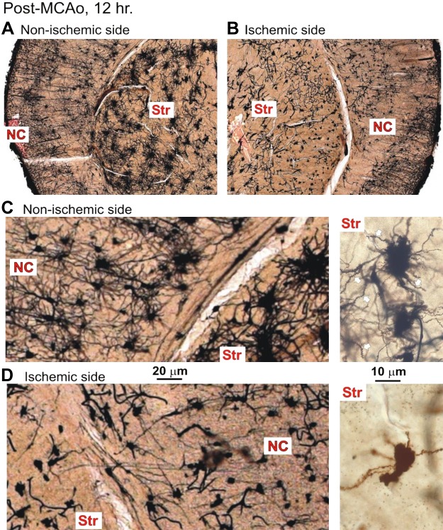
Fig. 11.
A: the nonischemic hemisphere contains neurons with extensive dendritic arbors, in both neocortex (NC) and striatum (Str). B: in contrast, the ischemic hemisphere shows punctate cell bodies and reduced dendritic branching in the middle cerebral artery occlusion (MCAo) core in both NC and Str. C: at higher magnification, pyramidal cells in the NC are abundant. The spider-like medium spiny neurons in Str also appear healthy, with short but numerous dendrites decorated with spines (white arrows; inset right). D: in contrast, the ischemic hemisphere is populated with dysmorphic, necrotic cell bodies with stunted dendrites in both NC and Str. Some striatal neurons have lost most of their dendrites, whereas the remainder have stunted, beaded dendrites devoid of spines (inset right). MCAo, middle cerebral artery occlusion.

