
Fig. 6.
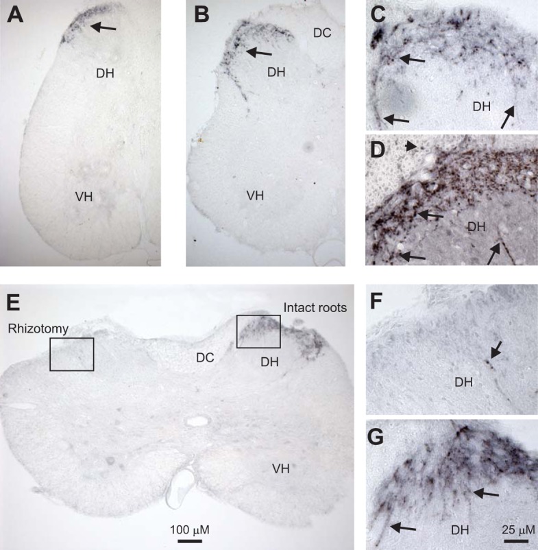
5-HT1D receptors are mostly expressed on small sensory afferents in the superficial dorsal horn. A: 5-HT1D receptor immunolabeling (black, diaminobenzidine) in sacral spinal cord of a normal rat. B: similar immunolabeling in a chronic spinal rat, caudal to injury. Note immunolabeling is only in the superficial dorsal horn (DH; at arrows). Also, labeling is absent from the dorsal columns (DC), where large propriospinal axons travel, and mostly absent from the ventral horn (VH), where propriospinal afferents synapse onto motoneurons in the monosynaptic stretch reflex (MSR) pathway. Very weak labeling of small γ-motoneurons was sometimes seen in the VH (see also E). C and D: magnified immunolabeling in the DH of normal and injured rats, respectively, showing labeling on fine beaded fibers confined mostly to the superficial laminae (arrows). Labeling was also seen on fine fibers in the dorsal root (arrowhead in D), consistent with the 5-HT1D labeling arising from fine sensory C-fibers. E: cutting the dorsal roots on the left side (rhizotomy) 4 wk before tissue fixation eliminated most labeling, indicating that the 5-HT1D receptors were mostly on fine sensory afferents. F and G: magnified images of the DH on the left and right sides (boxes) in E, showing only one weakly labeled fiber after rhizotomy. Scales for low- and high-magnification images are shown in E and G, respectively; n = 5 animals per condition, all showing the same results.