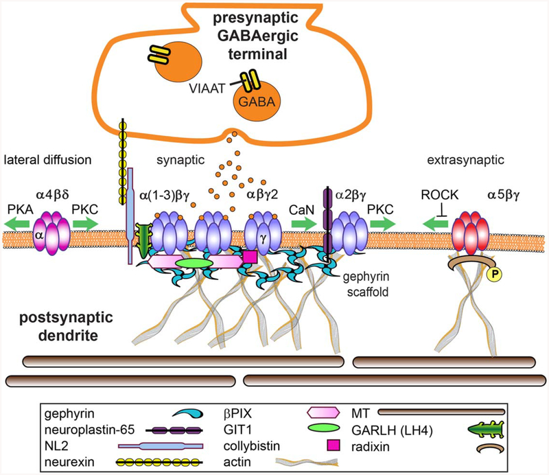
Figure 3.
GABAAR synapse structure. GABAARs composed of α(1–3)βγ subunits are largely syn-aptically localized via gephyrin interactions and contribute to phasic currents, whereas α(4 or 6)βd receptors are extrasynaptic and generate tonic current. α5βγ receptors are found in both locations due to binding with gephyrin at synapses and radixin extrasynaptically. Proteomics and other mod ern strategies have significantly enriched the complexity of the inhibitory synapse, however the functions of many new components have yet to be defined. Key synaptic adhesion, scaffold and sig naling proteins that mediate lateral diffusion of receptors in and out of the synapse are depicted and discussed in this review. [Color figure can be viewed at wileyonlinelibrary.com]

