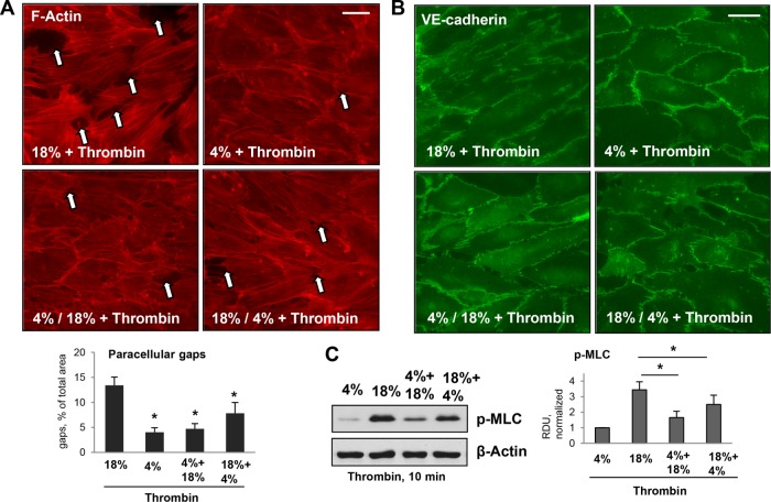
FIGURE 2:
Effects of different patterns of applied CS on thrombin-induced cytoskeletal remodeling and MLC phosphorylation in human pulmonary ECs. Pulmonary ECs were treated by 18% CS or 4% CS for 6 h; alternatively, 3-h 4% CS was introduced either before or after 3-h 18% CS. Thrombin (0.05 U/ml) was added in the last 10 min of CS stimulation. (A) Visualization of F-actin using immunofluorescence staining with Texas Red phalloidin. Paracellular gaps are marked by arrows. Bar = 10 µm. Bar graphs represent quantitative analysis of gap formation. Data are expressed as mean ± SD; n = 3; *, p < 0.05. (B) Visualization of cell junctions using immunofluorescence staining with VE-cadherin antibody. Bar = 10 µm. (C) Thrombin-induced MLC phosphorylation in pulmonary ECs exposed to different patterns of CS was evaluated by Western blot with pp-MLC antibodies. Equal protein loading was confirmed by membrane reprobing with β-actin antibody. Bar graphs represent analysis of Western blot data; n = 4; *, p < 0.05.

