FIGURE 5:
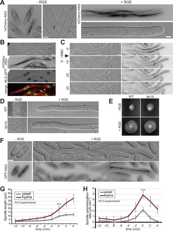
Microtubules are not involved in hyphal growth. (A) Microtubule organization in S. japonicus yeast and hyphal forms expressing mCherry-Atb2. Images are maximum-intensity projections of 8–14 z-stacks. (B) Images of an induced strain tagged with mCherry-Atb2 and NLS-GFP showing that microtubules can penetrate the space between the plasma membrane and the vacuole (arrowhead). (C) Microtubule depolymerization does not perturb hyphal growth. MBC was added at time 0 in a microfluidic chamber. (D) Wild-type and tip1∆ cells grown in microfluidic chambers under inducing and noninducing conditions. (E) Wild-type and tip1∆ strains grown on solid media under noninducing and inducing conditions. Dotted lines highlight penetrative filamentous growth. (F) Mitotic spindles labeled with GFP-Atb2 in yeast and hypha. (G) Quantification of spindle length over time, aligned on the steepest slope and averaged (n = 30 cells per cell type). (H) Quantification of spindle elongation rates over time. Individual profiles were aligned on the highest rate and averaged (n = 30 cells per cell type). *** indicates p < 1.59 × 10⁻¹⁰; t test. Error bars show standard deviations. Scale bars: 5 µm.

