
Figure 1.
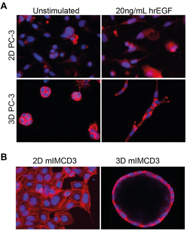
3D cell cultures provide a more physiologically relevant context for drug screening. (A) Prostate carcinoma (PC-3) cells cultured as 2D monolayer (top) show negligible morphological changes in response to growth factor (20 ng/mL hrEGF) stimulation but become invasive if embedded in 3D hydrogels (bottom) after growth factor stimulation. These invasive characteristics can be used to investigate the efficacy of inhibitors of receptor tyrosine kinases.96 Images in the top panel were obtained using a wide-field BD pathway 855 with a 10× objective, and images in the bottom panel were obtained using a Nikon Ti Eclipse confocal microscope with a 20× objective. (B) mIMCD3 cells transduced with a short-hairpin targeting Pkd1 form a monolayer in 2D culture (left panel, BD pathway 855 with 10× objective), but form cysts in 3D hydrogels, representing a more pathophysiologically relevant model of PKD (right panel, Nikon Ti Eclipse confocal microscope with 20× objective).108 F-actin (rhodamine-phalloidin), red; nuclei (Hoechst 33258), blue.