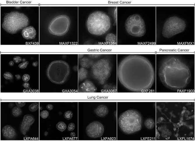
Figure 3.
3D cultures of PDX material. PDX material from different tumors can be cultured in 3D hydrogels to form complex microtissues that can be used for compound screening in a preclinically relevant context. Actin cytoskeleton visualized with rhodamine-phalloidin. PDX tumor material provided by Charles River Labs (Freiburg, Germany). Annotations refer to tumor type and PDX model number; BX = bladder; MAX = mammary; GX = gastric; PAX = pancreatic; LX = lung. 3D cultures and images generated by OcellO B.V.

