Figure 6.
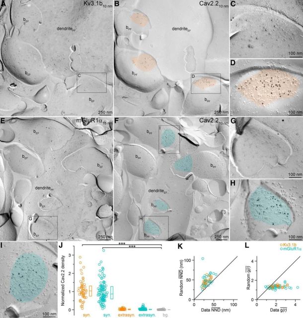
The densities of Cav2.2 subunit in AZs of PC boutons targeting Kv3.1b+ and mGluR1α+ dendrites. A, B, A Kv3.1b+ dendrite (dendritePF in A) is contacted by boutons with PF (bPF) membranes that show strong Cav2.2 subunit reactivity in their AZs (orange in B). C, D, High-magnification images of the boxed areas shown in A and B. E, F, PF bouton membranes contacting an mGluR1α+ dendrite (dendritePF in E) contain Cav2.2 subunit immunolabeled AZs (cyan in F). G–I, Enlarged views of the boxed areas in E and F. J, Normalized densities of gold particles labeling the Cav2.2 subunit within presynaptic AZs (syn.) and extrasynaptic membranes (extrasyn.) of boutons contacting Kv3.1b+ or mGluR1α+ profiles and in surrounding EF membranes (background: bg., gray). Post hoc MW U tests with Bonferroni correction after Kruskal–Wallis test (p < 0.0001) demonstrated a significant difference between the synaptic and background labeling (p < 0.0001). Circles indicate individual measurements of AZs, boxes indicate IQRs, and horizontal bars indicate medians. K, Comparison of NND's measured in 21 Kv3.1b+ and 40 mGluR1α+ dendrite-contacting AZs to their random controls (repeated 1000 times). Wilcoxon signed-rank test revealed significant difference (p < 0.0001) between the data and the random distributions for both AZ populations. L, g(r) of individual Kv3.1b+ (n = 21) or mGluR1α+ (n = 40) dendrite-targeting AZs is plotted against the g(r) of their random controls (repeated 1000 times). Wilcoxon signed-rank test revealed significant differences (p < 0.0001) between the data and the random distributions for both AZ populations.

