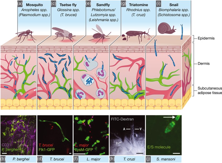
Figure 1.

Anatomically, the skin is divided into three compartments: the epidermis (an avascular layer mostly composed of keratinocytes and Langerhans cells), the dermis (highly perfused by blood [red] and draining lymph vessels [green]). (a) Plasmodium parasites are transmitted by infected Anopheles mosquitoes. Inoculated Plasmodium sporozoites can either remain in the skin, be transported to lymph nodes via lymph, or to the liver via blood vessels. Sporozoites show different migration dynamics at different skin sites or when in proximity to blood capillaries. (b) Maximum projection of P. berghei sporozoite tracks (green) in the proximity of blood vessels (CD31, magenta). Scale bar = 25 μm (Hopp et al., 2015). (c) Trypanosoma brucei parasites are transmitted by the bite of infected tsetse flies (Glossina spp.). They first develop in the skin at the bite site before they reach the blood and lymph vasculatures, and the entire dermis is an important parasite reservoir. (d) Extravascular trypanosomes (red) imaged in the skin vessels of Flk1‐GFP mice (green) using spinning‐disc confocal microscopy. Scale bar: 10 μm (Capewell et al., 2016). (e) Leishmania spp. are transmitted by the bite of infected Phlebotomine or Lutzomyia sandflies. Neutrophils are recruited to the bite site and are crucial for the dissemination of parasites. (f) Two‐photon IVM still from a LysM‐GFP mouse (neutrophils, green) 2 hr after infection with Leishmania major (red). Scale bar = 20 μm (Peters et al., 2008). (g) Trypanosoma cruzi is transmitted via the faeces of the triatomine Rhodnius prolixus. (h) Vascular permeability as shown by FITC‐Dextran leakage from the blood vessel upon insect probing. Scale bar = 100 μm (Soares et al., 2014). (i) Schistosoma spp. undergo asexual reproduction in freshwater snails (Biomphalaria spp.). Schistosoma spp. cercariae are motile and invade the host skin. (j) Schistosoma excretory/secretory molecule release (using a fluorescent tracer [green]) during parasite skin invasion. Scale bar = 100 μm (Paveley et al., 2009)
