
Fig. 6.
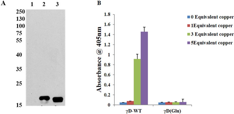
Effect of Cu2+ ions in the aggregation of glutathionylated γD crystallin WT. (A) Western Blot analysis of hγD incubated with GSSG using monoclonal GSH antibody. Antibody and the blot was developed using X-ray film using ECL solution the developed film was scanned under greyscale using Epson scanner 1: γD crystallin WT alone, 2 & 3: γD crystallin reacts with GSSG as indicated by the appearance of an immunoreactivity band. (B) Turbidity assays (n = 6) of hγD crystallin, glutathionylated (Glu) hγD (50 μM) incubated without or with 1, 3 and 5 equivalent of Cu2+ for 60 min.