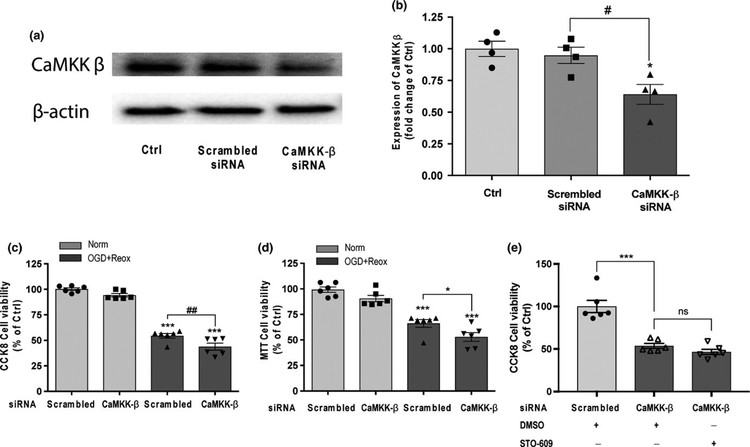
FIGURE 2.
Knockdown of CaMKK β expression exacerbated cell death under OGD with reoxygenation conditions in HBEC-5i endothelial cells. (a) and (b) Representative western blot images and quantitation of CaMKK β expression in HBEC-5i treated with scrambled siRNA and CaMKK β siRNA for 2 hr under normoxic conditions. Cells without siRNA treatment were considered control cells (Ctrl). Quantitation of CaMKK β was normalized to β-actin levels. (c) and (d) CaMKK β knock down using siRNA reduced cell viability in HBEC-5i cells under 18-hr OGD with 24 hr of reoxygenation (OGD + Reox) in the CCK-8 cell proliferation assay and MTT reduction assay. Scrambled siRNA (60 nM) and CaMKK β siRNA (60 nM) were incubated with the cells for 7 hr, normal growth medium containing 2× normal serum and antibiotic concentration was added and incubated for 24 hr. Cells were replaced with fresh 1× normal growth medium for another 24 hr of recovery prior to OGD and reoxygenation treatments. (e) STO-609 and CaMKK β had no additive effect in cell viability after OGD using CCK-8 cell assay. CaMKK β siRNA and STO-609 (10 μM) were added prior to OGD (18 hr) and maintained during reoxygenation (24 hr). Cells treated with scrambled siRNA under normoxic conditions (Norm) were considered control cells (Ctrl). Data are expressed as the means ± SEM of at least four independent experiments. *p < 0.05 and ***p < 0.001 versus Ctrl in the corresponding experiment. #p < 0.05 and ##p < 0.01 between the indicated treatments. Statistical analyses were performed using one-way ANOVA and the Holm-Sidak multiple comparison test

