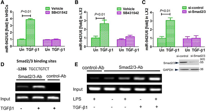
Fig. 3.
Smad2/3 mediate the induction of miR-942 expression in response to TGF-β. a Real-time PCR showing miR-942 levels in hHSCs and b in LX2 cells after 24 h stimulation with 5 ng/ml TGF-β1. The cells were pre-treated for 2 h with 10 μM SB431542 (a TGF-β1 receptor inhibitor). Un untreated. Bars show mean ± SD, n = 3 per group, of three independent experiments. c miR-942 levels as measured after 48 h of Smad2/3 knockdown in LX2 cells, and following stimulation with 5 ng/ml TGF-β1 for 24 h. Un untreated. Data represent mean ± SD of three independent experiments, n = 3 per group. One-way ANOVA (and nonparametric) Kruskal–Wallis test was used for statistical comparison of data (a–c). d Predicted Smad2/3 binding site at position − 1286 of the miR-942 promoter as identified by bioinformatics analysis, and ChiP analysis showing the effect of TGF-β stimulation (5 ng/ml). e ChiP assay showing the combined effect of 100 ng/ml LPS and 5 ng/ml TGF-β stimulation (2 h) on the recruitment of Smad2/3 to Smad binding site in the hsa-miR-942 promoter in LX2 cells. Un untreated, Ab antibody. LX2 cells pre-treated with LPS for 2 h in the combination treatment. Then, cells were lysed, nuclei were purified and soluble chromatin was immunoprecipitated using anti-Smad2/3 or normal rabbit IgG as control Ab. Subsequently, the miR-942 promoter fragment containing the – 1286-binding site was amplified by PCR

