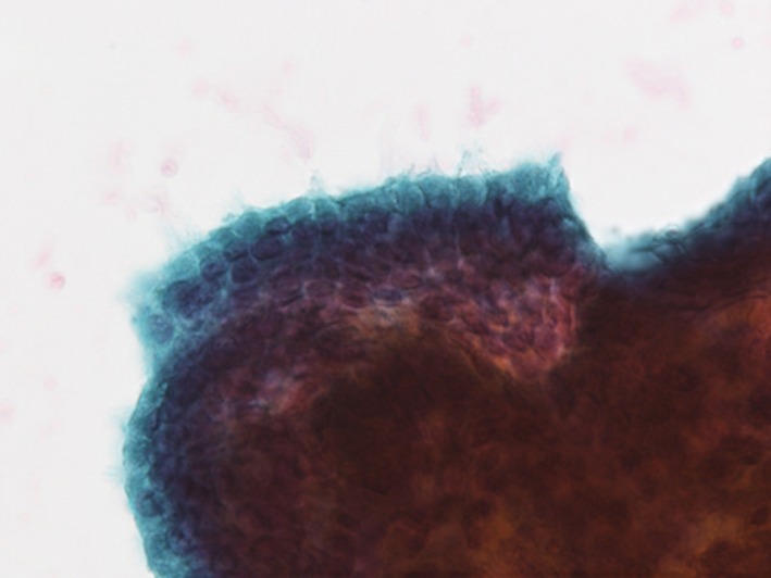
Figure 2.

A palisaded arrangement composed of tall columnar carcinoma cells is seen at the periphery of large cell nest. The nuclei are overlapped and located on the basal side of the carcinoma cells. (Papanicolaou stain, 200×) [Color figure can be viewed at wileyonlinelibrary.com]
