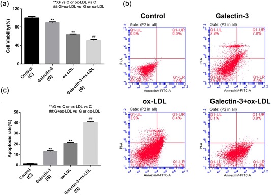
Figure 2.

The effects of Gal‐3 on cell viability and apoptosis in ox‐LDL‐treated HUVECs. Cells were treated with 250 ng/ml Gal‐3 for 48 hr followed by treatment with ox‐LDL for 6 hr. (a) Cell viability was measured by the MTT assay. (b) Apoptosis was assessed by flow cytometry. (c) The rate of apoptosis was indicated using histograms. Each experiment was performed in triplicate. **p < 0.01, ## p < 0.01 [Color figure can be viewed at wileyonlinelibrary.com]
