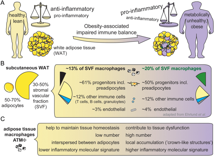
Figure 1.
Obesity-associated impaired immune balance in white adipose tissue. (A) Obesity is associated with an impaired immune balance toward pro-inflammatory in WAT. All fat depots are affected, but mostly the viscWAT. (B) ATMΦ amount is low in lean scWAT (~13% of SVF). However, MΦ are numerically the dominant type of immune cells representing half of the immune cells. MΦ increase in obese WAT, for example in human scWAT from 13 to 20% of the SVF (36). (C) The roles of ATMΦ in lean (left) and obese (right) WAT. The number of MΦ is low and they are interspersed between adipocytes in WAT of lean subjects, contrasting the higher number and local accumulation of MΦ in crown-like structures during obesity, which is fostered by proliferation, high immigration and low emigration. The low inflammatory profile (surface markers, cytokine expression and secretion, e.g. IL4, IL10) in lean subjects transforms into higher inflammatory status (e.g. TNFα, IL6, IL1β) during obesity.

 This work is licensed under a