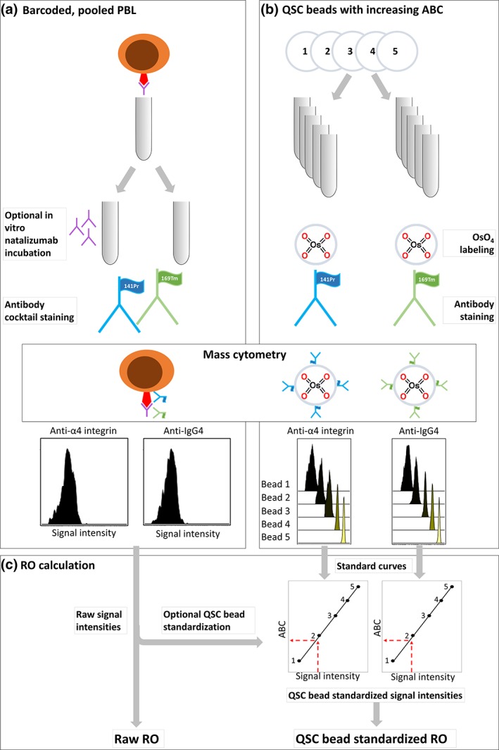
Figure 2.

Experimental workflow: (a) peripheral blood leukocytes (PBLs) were split into two aliquots for optional in vitro incubation with natalizumab, stained with an antibody cocktail containing anti‐IgG4 and anti‐α4 integrin, and analyzed on a Helios mass cytometer. (b) Quantum simply cellular (QSC) beads with known antibody binding capacity (ABC) were labeled with OsO4, stained with anti‐IgG4 or anti‐α4 integrin, and acquired on the same mass cytometer on the same day. (c) Standard curves were created based on anti‐IgG4 and anti‐α4 integrin signal intensities from QSC beads with known ABC, and signal intensities of the same antibodies from the PBL samples were plotted into the standard curves for standardization before RO calculation. [Color figure can be viewed at wileyonlinelibrary.com]
