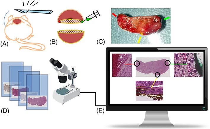
Figure 1.

Obtaining digital histology images for alignment with the corresponding MR parameter maps. A) The dead animal is positioned for incision through skin and tumour, with scalpel cutting adjacent and parallel to the imaged tumour plane. B) Colour coding of the imaged tumour plane by tissue ink injections on the left, right and dorsal tumour borders. C) Digital photography of the divided and colour coded tumour, with indications of where the ink is visible. The plane of the surface facing the camera will be parallel to the microtome knife sweep plane. D) Glass microscopy slides with differently stained parallel sections are digitized using a slide scanner. E) The ink, preserved through the preparation and paraffin embedding procedure, is visible on the magnified portions of the digitized HE stained section, and can be used to recover the orientation of the histological sections for alignment with the MR images
