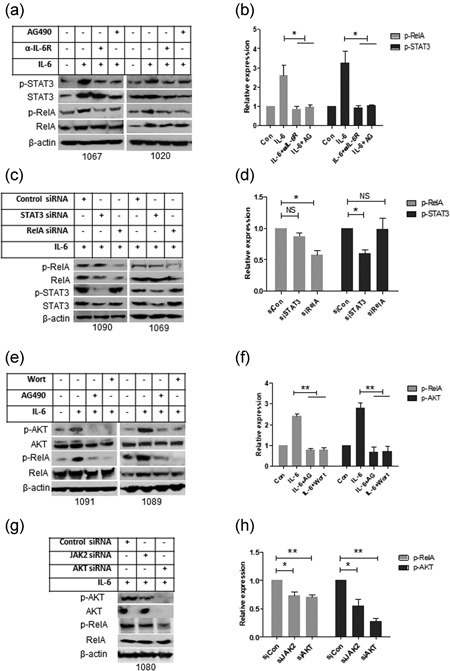
Figure 4.

Functional analysis of the IL‐6‐mediated signaling pathway. (a,b) After IL‐6 receptor was blocked by anti‐IL‐6 receptor antibody and JAK2 was blocked by AG490; primary CLL cells were incubated with IL‐6 for 16 hr. (c,d) STAT3 or RelA was knocked down by transfection of STAT3 or RelA siRNA, and cells were incubated with IL‐6. The levels of P‐STAT3 and P‐RelA were determined by western blot analysis. (e,f) CLL cells were treated with PI3K inhibitor Wortmannin or AG490 and then incubated with IL‐6 for 16 hr. (g,h) JAK2 or AKT was knocked down by transfection of JAK2 or AKT siRNA and after 24 hr cells were incubated with IL‐6 for 16 hr. Phosphorylation of AKT and RelA was determined by western blot analysis. A, C, E, and G are representative western blot analysis and B, D, F, and H are statistical analysis of at least three cases data. Relative expression of phosphorylated protein was analyzed by densitometry using actin as control. Data shown are mean ± SD from at least three independent experiments. CLL: chronic lymphocytic leukemia; IL‐6:interleukin 6; SD: standard deviation
