Figure 5.

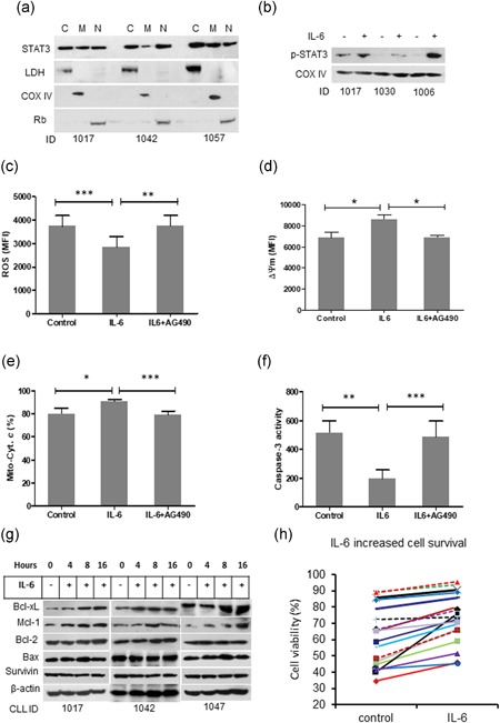
IL‐6 prevents mitochondria‐dependent apoptosis in CLL cells. (a) STAT3 intracellular distribution. Fresh primary CLL cells were fractionalized into cytoplasm (c), mitochondria (M) and nucleus (N). STAT3 expression was determined by western blot analysis. LDH, COX IV, and Rb are markers for cytoplasm, mitochondria and nucleus, respectively. (b) IL‐6 induced phosphorylation of STAT3. (c) ROS production, (d) Mitochondrial depolarization, and (e) cytochrome c release were measured by flow cytometry. (f) Caspase‐3 activity was measured by a fluorogenic assay. Data from A to F are mean ± SD from three independent experiments. Significant difference was analyzed by the Student t‐test. (g) Effect of IL‐6 on expression of Bcl‐xL, Mcl‐1, Bcl‐2, Bax, and Survivin was determined by western blot analysis. (h) CLL cell viability. CLL cells were incubated with or without IL‐6 for 24 hr. Cell viability was determined by flow cytometry after cells were stained with PI. CLL: chronic lymphocytic leukemia; IL‐6: interleukin 6; PI: propidium iodidie; ROS; reactive oxidative species; SD: standard deviation [Color figure can be viewed at wileyonlinelibrary.com]
