Figure 6.

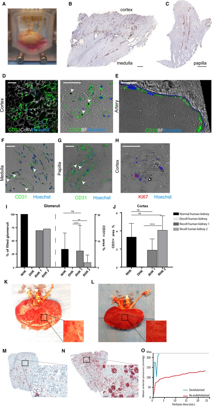
Human kidney re‐endothelialization with hiPS‐ECs. A, Image of the human re‐endothelialized kidney in the custom‐made organ chamber during culture. B, Representative immunohistochemistry images of CD31+ showing re‐endothelialization in the cortex, medulla, and (C) papilla. D, Representative CD31 immunofluorescent images show re‐endothelialization of the kidney cortex, (E) artery, (F) medulla, and (G) papilla. CD31 dynabeads used in the iPS‐EC differentiation protocol (white arrowheads) can still be observed. H, Representative image of proliferating cells in the human re‐endothelialized kidney. I, Quantification of CD31. For the recellularized human kidneys (N = 2) 4 areas per kidney were sampled and analyzed. Per slide all glomeruli were annotated manually (about 100 glomeruli per slide). The graphs display the amount of filled glomeruli as a % of all analyzed glomeruli per kidney (left) and the % of CD31+ area within all filled glomeruli (right) at 24 h after infusion of cells. ****P < .0001 **P < .01. J For cortex analysis %CD31+ area was calculated within whole cortex area per kidney. The graphs display the average of 4 different sample areas per kidney. ****P < .0001. K, Macroscopic image of a decellularized human kidney perfused with whole blood showing only some “patchy”perfused areas. L, Macroscopic image of a re‐endothelialized human kidney scaffold perfused with whole blood showing diffuse blood coverage over the whole kidney. M, Microscopic picture of a Movat's‐Pentachrome staining of a decellularized human kidney cortex scaffold perfused with whole blood shows only a few perfused areas. Because of low pressure and massive leakage (due to lack of endothelial cells), only a few red blood cells reached the glomeruli. N, When the human re‐endothelialized kidney scaffold was perfused with whole blood, high coverage of blood in the cortex was observed (shown in red), showing that perfusion of blood through the entire kidney was possible. O, Whereas the whole blood perfusion of the decellularized human kidney scaffold was stopped within 5 min because of extremely high perfusion pressures, the re‐endothelialized kidney could be perfused for more than 20 min. Scale bar B, C 500 μm; scale bar D, F, H 50 μm; scale bar E 25 μm. White arrowheads: CD31 dynabead, black/white arrowhead: proliferating cell. Scale bar M, N 200 μm. hiPS‐ECs, human inducible pluripotent stem cell–derived endothelial cells; ns, nonsignificant
