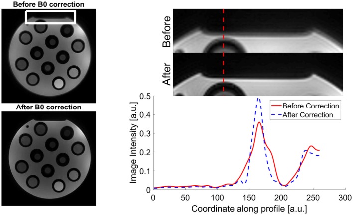
Figure 3.

Effects of deblurring using our 2‐step MRF reconstruction on a detail of our in vitro acquisition (white rectangle). As shown by the intensity profile along the dotted red line, the corrected image (dotted blue line) has sharper edges with respect to the original image (solid red line)
