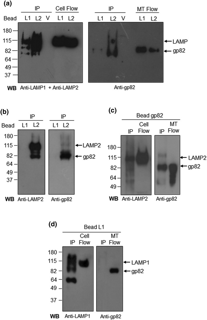
Figure 4.

Binding of metacyclic trypomastigote (MT) gp82 to LAMP‐2. (a) Protein A/G magnetic beads, cross‐linked to antibody to LAMP‐1 or LAMP‐2, were incubated for 1 hr with HeLa cell extracts and afterwards with MT lysates for 1 hr. The eluates corresponding to immunoprecipitates (IPs) were analysed by Western blot (WB). Shown are the IP from LAMP1 beads (L1), LAMP2 beads (L2), control void beads (V), and the flowthrough samples from L1 and L2 beads corresponding to HeLa cell extract (cell flow) or MT lysate (MT flow). The blots were revealed with a mix of antibodies to LAMP‐1 and LAMP‐2 or with anti‐gp82 antibody. Note that both LAMP‐2 and gp82 were detected in IP from L2 beads. (b) The assay described in (a) was repeated with a new batch of protein A/G magnetic beads cross‐linked to antibody to LAMP‐1 or LAMP‐2. Note the presence of LAMP‐2 and gp82 in IP from L2 beads. No bands were detected in IP from L1 beads. (c) Protein A/G magnetic beads, cross‐linked to anti‐gp82 monoclonal antibody, were incubated with MT lysate for 1 hr, followed by 1‐hr incubation with HeLa cell extract. The WB of IP revealed LAMP‐2 and gp82. Shown also are the flowthorough samples corresponding to HeLa cell extract (cell flow) and MT lysate (MT flow). (d) LAMP1 beads were incubated with HeLa cell extract for 1 hr, followed by 1‐hr incubation with MT lysate. The WB of IP was revealed with antibody to LAMP‐1 and gp82. Also shown are the cell flow and MT flow, which served as control. Note that gp82 is absent in IP from L1 beads
