
Figure 2.
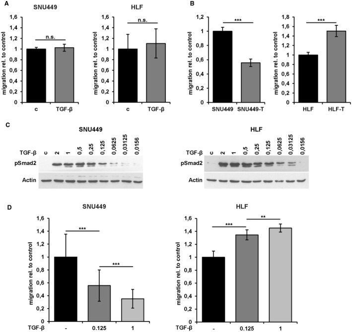
Duration‐dependent and concentration‐dependent migratory response of mesenchymal‐like HCC cells to TGF‐β treatment. (A) Migrated areas of HLF and SNU449 cells and those treated with 2.5 ng/mL TGF‐β1 for 24 hours in wound healing assays. (B) Migrated areas of SNU449 and HLF cells and those long‐term treated with 1 ng/mL TGF‐β1 (> 10 days, termed SNU449‐T and HLF‐T) in wound healing assays. (C) Western blot analysis of pSmad2 after long‐term treatment (> 10 days) of cells with different concentrations of TGF‐β1 (ng/mL). (D) Migrated areas of SNU449 cells (left panel) and HLF cells (right panel) after long‐term treatment (> 10 days) with 0.125 ng/mL and 1 ng/mL TGF‐β1. Data are expressed as mean ± SD. Error bars depict SD from at least three individual experiments. **P < 0.01, ***P < 0.001. Abbreviations: c, control; n.s., not significant.