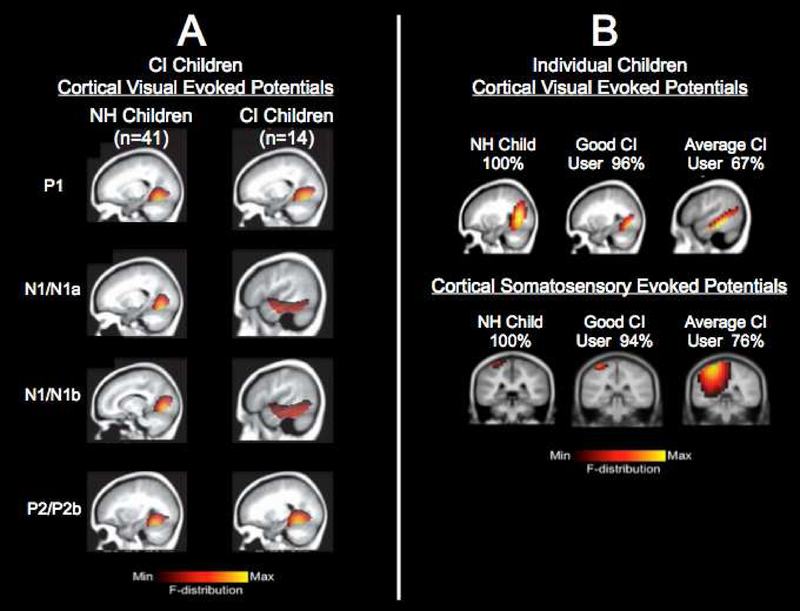
FIGURE 1. Visual Cross-Modal Plasticity in CI Children.
Panel 1A: Visual cross-modal re-organization in a group of CI children. Visual cortical evoked potential (CVEP) current density source for a group of normal hearing children (n=41) and a group of CI children (n=14) in response to a visual motion stimulus. For the higher-order CVEP components, cortical areas involved in processing of visual motion stimuli are observed for the normal hearing group, whereas the CI children show additional recruitment of temporal cortex. Adapted from Campbell & Sharma (2016).
Panel 1B: Visual cross-modal re-organization in individual CI children. Top Panel: Current density source reconstructions for the P2 cortical visual evoked potential (CVEP) component in a normal hearing child, a cochlear implanted child with excellent speech perception, and a CI child with average speech perception recorded in response to a visual motion stimulus. The normal hearing child and CI child with good speech perception show expected activation of cortical areas involved in processing of visual motion stimuli, while the CI child with average speech perception shows additional recruitment of temporal cortices. Bottom Panel: Current density source reconstructions for the N70 cortical somatosensory evoked potential (CSSEP) component in a normal hearing child, a CI child with excellent speech perception, and a CI child with average speech perception recorded in response to a 250 Hz tone applied to the right index finger. Vibrotactile stimulation in the normal hearing and CI child with excellent speech perception show expected activation of cortical areas involved in somatosensory processing, while the CI child with average speech perception shows additional recruitment of auditory cortex. Adapted from Sharma, Campbell, & Cardon (2015).

