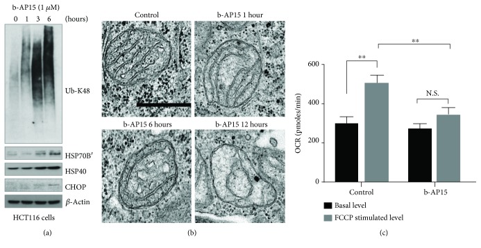
Figure 1.
Induction of mitochondrial dysfunction in HCT116 cells by the deubiquitinase inhibitor b-AP15. (a) HCT116 cells were exposed to 0.5% DMSO or 1 μM b-AP15 for 1, 3, and 6 hours, and extracts were prepared and subjected to immunoblotting using the indicated antibodies. (b) Electron micrographs of HCT116 cells treated with b-AP15 for 1, 6, and 12 h. Scale bar = 0.5 μm. (c) Basal and maximal oxygen consumption rates (OCR) were measured after a 5-hour exposure of HCT116 cells to 1 μM b-AP15 using a Seahorse XF analyzer. Uncoupled respiration was measured after exposure to carbonyl cyanide-4-(trifluoromethoxy)-phenylhydrazone (FCCP) (mean ± S.D.; ∗∗p < 0.01).

