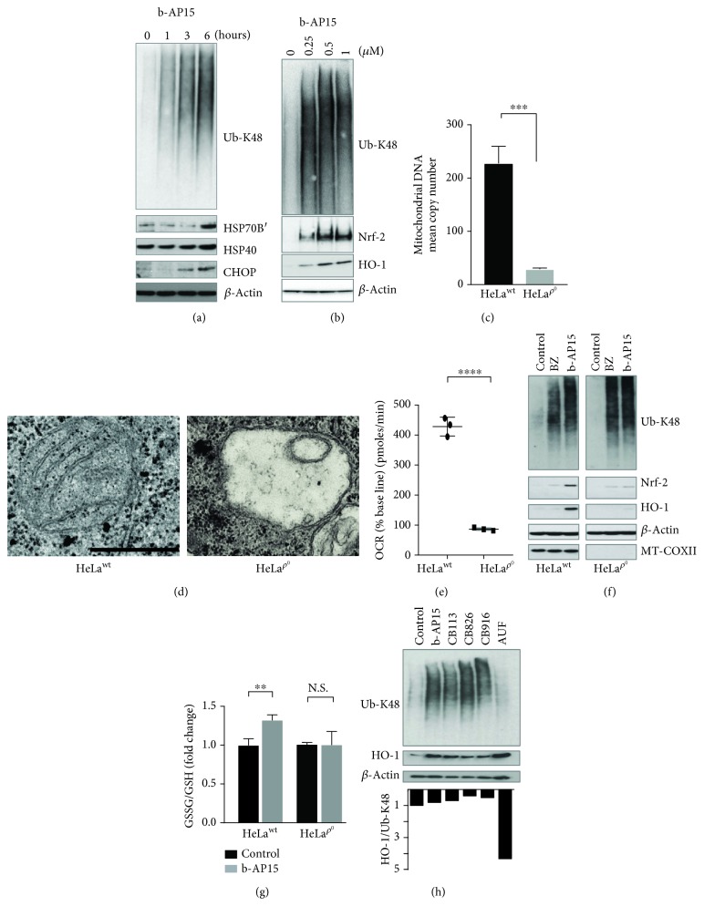
Figure 3.
HeLa Rho0 (ρ 0) cells show a decreased oxidative stress response to b-AP15. (a) HeLa cells were exposed to 0.5% DMSO or 1 μM b-AP15 for 1, 3, and 6 hours, and extracts were prepared and subjected to immunoblotting using the indicated antibodies. All cultures received 0.5% DMSO. (b) HeLa cells were exposed to 0.5% DMSO or b-AP15 (0.25, 0.5, and 1.0 μM in 0.5% DMSO) for 6 h, and extracts were prepared and subjected to immunoblotting using the indicated antibodies. (c) HeLa cells were exposed to EtBr and uridine to generate mitochondrial DNA depleted cells (HeLa ρ 0). The ratio of mtDNA to nDNA was compared in HeLa parental and ρ 0 cells using RT-PCR (∗∗∗p < 0.001). (d) Electron micrographs of mitochondria in HeLa parental and ρ 0 cells. Scale bar = 0.5 μm. (e) Basal oxygen consumption rates (OCR) of HeLa parental and ρ 0 cells (n = 3; mean ± S.D.; ∗∗∗∗p < 0.0001). (f) HeLa ρ 0 cells were treated with 100 nM bortezomib (BZ) or 1μM b-AP15 for 5 h followed by western blot analysis for K48-linked polyubiquitin chains, Nrf-2, HO-1, MT-COXII, and β-actin. Note the impaired induction of Nrf-2 and HO-1 by UPS inhibitors in ρ 0 cells. (g) The ratio of GSSG/GSH was determined in parental HeLa and ρ 0 cells exposed to 1 μM b-AP15 or vehicle for 6 h (n = 3; mean ± S.D.; ∗∗p < 0.01). (h) HCT116 cells were exposed to 0.5% DMSO, 1 μM b-AP15, 5 μM CB113, 5 μM CB826, 5 μM CB916, and 1.5 μM auranofin (AUF) for 6 h, and extracts were prepared and subjected to immunoblotting using the indicated antibodies.

