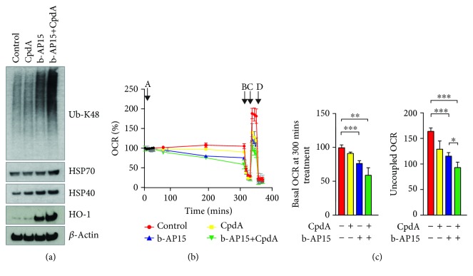
Figure 4.
Increased levels of proteotoxic stress are associated with decreased mitochondrial function and increased induction of HO-1. (a) HCT116 cells were exposed to 0.5% DMSO, 1 μM b-AP15, and 10 μM CpdA for 6 h, as indicated. Extracts were prepared and subjected to immunoblotting using the indicated antibodies. Note the increased levels of polyubiquitinated proteins, Hsp70, and HO-1 in cells exposed to b-AP15 and the ER translocation inhibitor CpdA. (b, c) HCT116 cells were treated with b-AP15 (1 μM) and/or CpdA (10 μM) for 5 hours and oxygen consumption rates were measured using a Seahorse XF analyzer (n = 3 in each group). A: DMSO or compounds; B: oligomycin; C: FCCP; D: antimycin and rotenone. (b) Measurement of OCR in real time after exposure to different compounds; (c) left: basal OCR after 300 min of treatment with compounds (mean ± S.D.; ∗∗∗p < 0.0001; n = 3); right: uncoupled OCR after addition of FCCP (mean ± S.D.; ∗∗∗p < 0.0001; ∗p < 0.05; n = 3).

