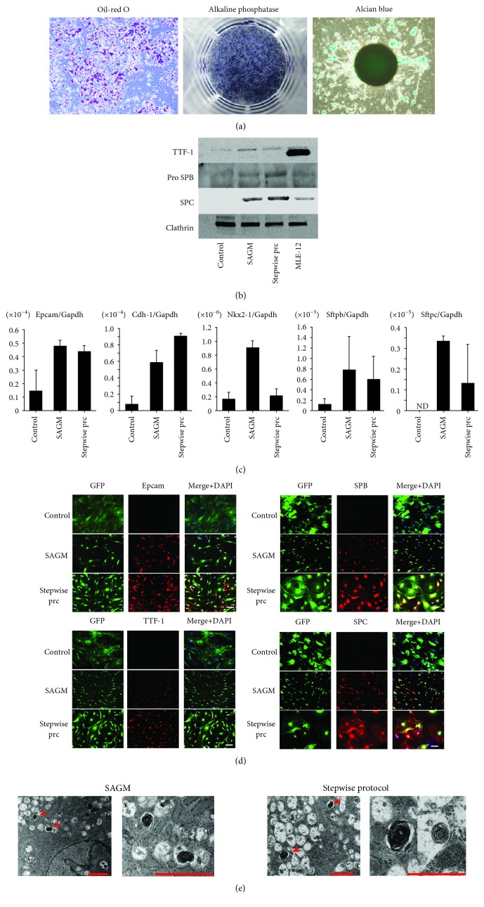
Figure 1.
Differentiation into trilineage cells and alveolar epithelial cells of adipose-derived stem cells (ADSCs). (a) Histological analysis of differentiation of ADSCs into adipogenic, osteogenic, and chondrogenic lineages. Adipogenic differentiation was assessed with oil red O staining. Osteogenic differentiation was examined using alkaline phosphatase staining. Chondrogenic differentiation of ADSCs was examined by alcian blue staining. ADSCs have the capacity to differentiate into each mesenchymal lineage. Scale bars: 50 μm. (b) Western blot analysis of ADSCs. Expression of TTF-1, Pro SPB, and SPC is shown. MLE-12 is a murine lung epithelial cell line. (c) Real-time quantitative RT-PCR analysis of ADSCs. ADSCs were cultured in small airway growth medium (SAGM) or with the stepwise protocol for 28 days. Expression of EPCAM and Cdh-1 (epithelial markers) and Nkx2-1, Sftpb, and Sftpc (type 2 alveolar epithelial cell markers) was analyzed with RT-PCR on day 28 (n = 3). Each value was normalized to the level of Gapdh. ND: not detected. (d) Immunofluorescence staining of ADSCs with anti-EPCAM, anti-TTF-1, anti-SPB, and anti-SPC. Control: ADSCs cultured in growth medium; SAGM: ADSCs cultured in SAGM; stepwise prc: ADSCs cultured with the stepwise protocol. Scale bars: 100 μm. (e) Transmission electron microscopy of ADSCs. Lamellar body-like structures were observed in ADSCs cultured in SAGM or with the stepwise protocol for 28 days. Scale bars: 2 μm.

