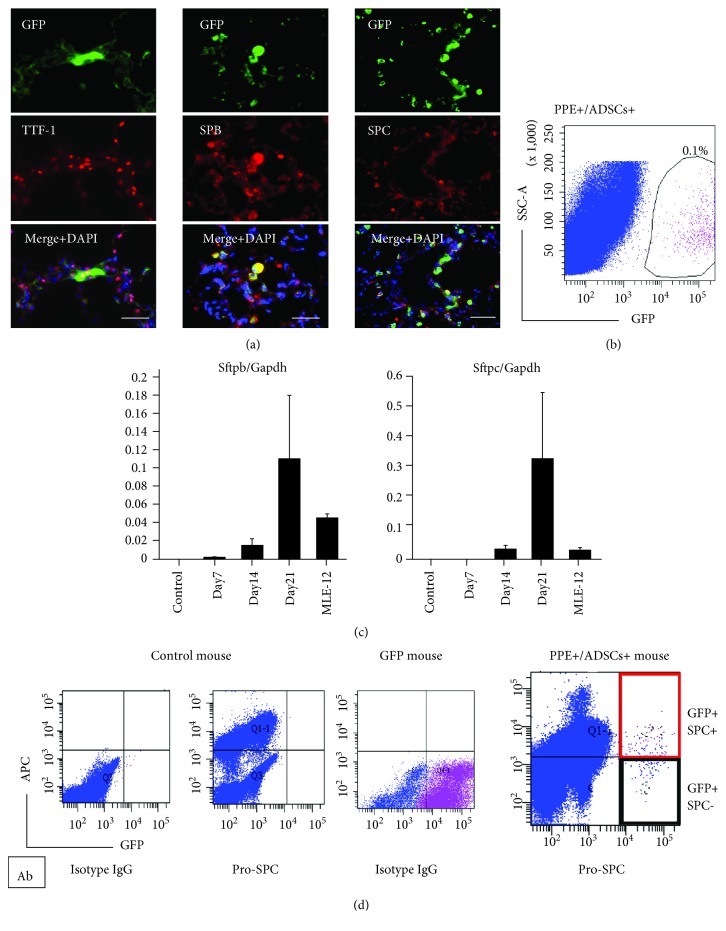
Figure 3.
Differentiation of engrafted GFP-labeled ADSCs in the lung of the emphysematous mouse model. (a) Immunohistochemical staining of anti-TTF-1, anti-SPB, and anti-SPC in GFP-positive cells in an emphysematous mouse lung. Scale bars: 50 μm. (b) Isolation of GFP-labeled ADSCs with fluorescence-activated cell sorting (FACS) on day 21. (c) Real-time quantitative RT-PCR analyses of Sftpb and Sftpc in GFP-labeled ADSCs sorted from a murine lung with FACS (each day, n = 6). (d) Cells were isolated from the lungs of control mice, GFP mice, and PPE+/ADSCs+ mice and analyzed for the percent of GFP+/SPC+ cells by flow cytometry. A representative FACS dot plot is shown.

