Figure 4.
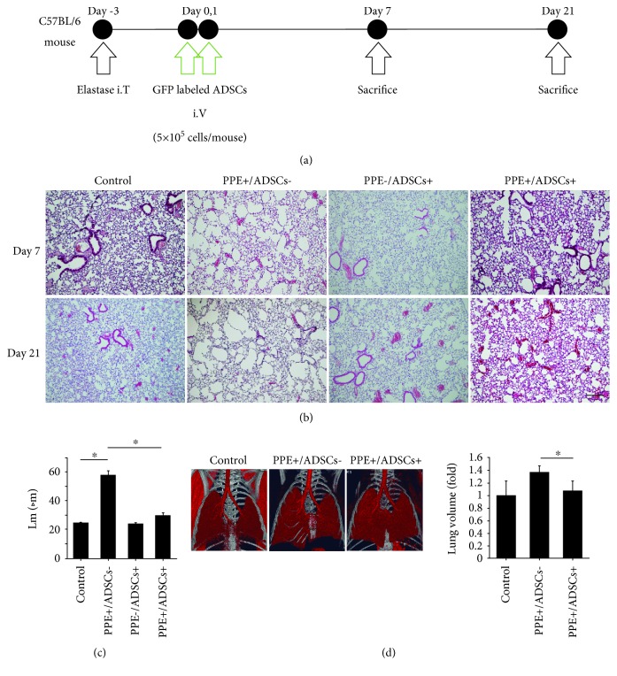
Histological effects of the administration of ADSCs in emphysema model mice. (a) The timeline of experiments. Emphysema was induced in C57BL/6 mice by intratracheal administration of elastase (porcine pancreatic elastase (PPE), 1.5 IU/mouse). After 3 days, ADSCs were intravenously administered (5 × 10⁵ cells/mouse) on days 0 and 1. Their effects were evaluated histologically on days 7 and 21. (b) Representative histological images on days 7 and 21 with hematoxylin and eosin staining. ADSCs ameliorated emphysematous changes. Scale bars, 200 μm. (c) The mean linear intercepts (Lm) on day 21. Airspace enlargement was quantified by measuring the Lm (n = 3). ∗ P < 0.05. (d) Representative three-dimensional CT images. The lung volumes were measured using the product of the maximum length in the craniocaudal and lateral axes on day 21 on three-dimensional CT images (n = 4 in the control group, PPE+/ADSC− group, and PPE+/ADSC+ group on each day).

