
Figure 3.
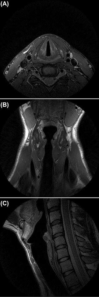
High‐resolution T2 FSE weighted (PROPELLER) axial (A), coronal (B), and sagittal (C) images of the larynx of a healthy volunteer. Pediatric laryngeal MRI protocol developed at the Erasmus MC—Sophia Children's Hospital, with the use of a 3T MRI (GE Healthcare) using a 6 Chanel Carotid coil (spatial resolution 0.5x.0.5 (in plane) x 2 mm).