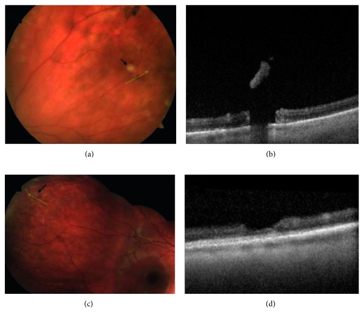
Figure 2.
Peripheral SD-OCT of operculated breaks and focal operculated schisis. (a) Peripheral photograph of the left eye of this asymptomatic patient with an operculated break; black arrow denotes operculum; green line scan demonstrates scan position through the break. (b) SD-OCT reveals a full-thickness retinal break. (c) Photograph of the right eye of this symptomatic patient with an apparent full-thickness operculated break. (d) SD-OCT reveals focal operculated schisis with no full-thickness component.

