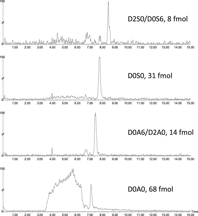
Fig. 4.
Extracted ion chromatograms of heparan sulfate disaccharides extracted from the surface of a grade 2–3 prostate cancer tissue microarray core following on surface heparinase I-III digestion. The amount of the extracted disaccharides were estimated based on the calibration curve obtained using the standards.

