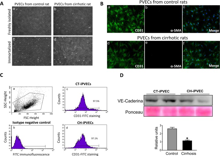
Fig 1. Immortalized PVEC maintain endothelial cell features.
PVECs were isolated from portal veins from control and cirrhotic rats. Immortalization of the cells was carried out using a retrovirus long sequence containing the SV40 virus T antigen. A Images taken by optical microscope of PVECs from control (left) and cirrhotic (right) rats, before (top) and after (bottom) immortalization, are shown. B CD31 and α-SMA immunostainings are shown for iPVECS established from control (a, b and c panels) and cirrhotic rats (d, e and f panels). The merged panels show CD31 and α-SMA colocalization. Nuclei were stained with DAPI (blue) (n = 5). Original magnification 200X. C iPVECs were immunostained for CD31 and analyzed by flow cytometer. Panel a shows dot-blot graph of the cell population. The negative population for the CD31 antigen was chosen from cells quantified in the absence of anti-CD31 antibody (panel c), CT-iPVEC (panel b), and CH-iPVEC (panel d) (n = 5). D cell lysates were analyzed by western blot. Upper panel shows VE-Cadherin immunoblot with loading control by ponceau staining. Lower panel shows densitometric analysis of the western blot. *p<0.05 vs control.

