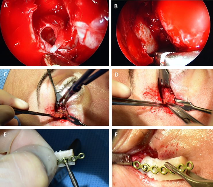
Fig 1. All patients underwent balanced orbital decompression.
The extent of surgery was chosen according to the individual proptosis. (A-B) The medial wall was decompressed via endonasal endoscopy. The periorbit was incised allowing the orbital content to prolapse into the ethmoid cavity. (C-D) Resection of a bony triangle of the lateral wall was performed through a 10mm incision next to the lateral canthus. (E-F) To restore the lateral orbital rim, the anterior part of the resected bone is replanted employing a fixation with microplates.

