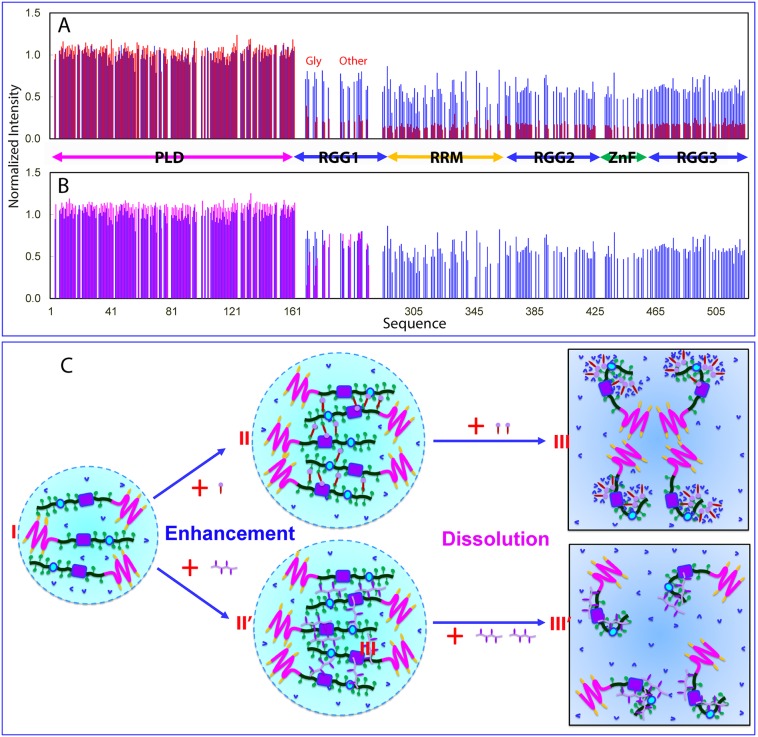
Fig 8. NMR view of enhancement and dissolution of LLPS of FUS induced by ATP and ssDNA.
(A) Normalized HSQC peak intensity of the 15N-labeled FUS in the presence of TssDNA at molar ratios of 1: 0.1 (blue) and 1:0.5 (red) as divided by that of FUS in the free state. (B) Normalized HSQC peak intensity of the 15N-labeled FUS in the presence of TssDNA at molar ratios of 1: 0.1 (blue) and 1:10 (purple) as divided by that of FUS in the free state. (C) A speculative model to rationalize the specific binding of ATP and ssDNA to Arg/Lys residues as well as RRM and ZnF of FUS to enhance LLPS at low concentrations but dissolution at high concentrations. FUS, Fused in sarcoma; HSQC, Heteronuclear single quantum coherence spectroscopy; LLPS, liquid–liquid phase separation; PLD, prion-like domain; RGG, Arg-Gly/Arg-Gly-Gly–rich region; RRM, RNA-recognition motif; ssDNA, single-stranded DNA; TssDNA, telomeric ssDNA; ZnF, zinc finger.

