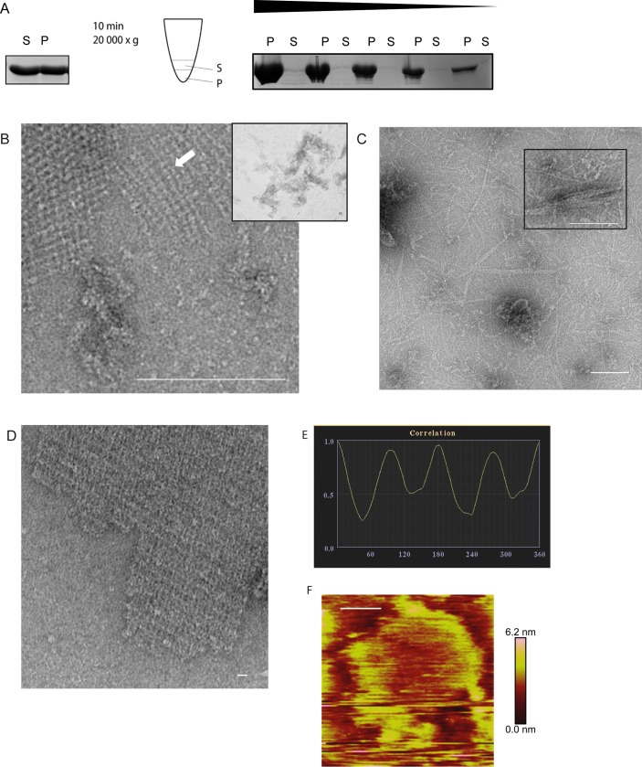
Fig 5. Ultrastructure of HP1542.
(A) Spin-down assay of HP1542 after purification from E. coli. Left: coomassie-stained SDS-PAGE of the spin-down assay of freshly purified Strep-HP1542. Middle: pictogram demonstrating the procedure. Samples were split in two equal fractions: the upper one was defined as soluble fraction (S) and the remaining one was defined as pellet fraction (P). Right: coomassie-stained SDS-PAGE of the spin-down assays with decreasing protein concentrations of aggregated Strep-HP1542; (B-D) Transmission electron microscope (TEM) images of purified Strep-HP1542 in high (B, large image) and low magnification (B, small image), of purified HP1542-His (C) and of purified HP1542 without tag (D). (E) Example of symmetry determination of a unit cells via correlation averaging using the ANIMETRA software. (F) Atomic Force microscopy (AFM) topographic image of HP1542 assembled on mica. A height scale bar is shown to the right. Scale bars of all microscopic images are 100 nm.

