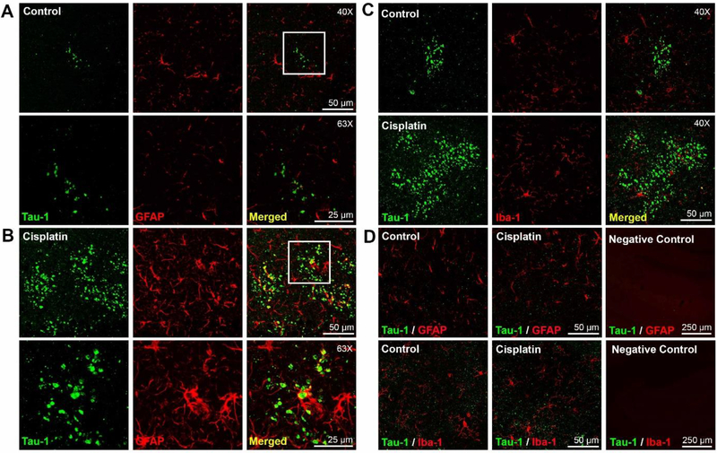
Figure 2. Accelerated development of tau clusters in cisplatin treated mice is associated with increased GFAP expression.
(A and B): Brains of control and cisplatin treated mice were co-stained for Tau-1 and GFAP. Representative examples of GFAP staining in an area of minimal Tau-1 clusters in control mice (A) and overt Tau-1 pathology in cisplatin treated mice (B). (Top row; 40x objective; Bottom row; 63x objective). The higher magnification demonstrates that there is no overlap between Tau-1 clusters and GFAP. C. GFAP staining in areas without Tau-1 clusters (left and middle panel) and control staining with secondary antibodies only (right). C. Double immunofluorescence analysis of Tau-1 and Iba-1 in brain of control (top) and cisplatin-treated (bottom) mice in areas of Tau-1 clusters. D. GFAP (top) and Iba-1 (bottom) expression in brain areas without Tau-1 clusters in control (left) and cisplatintreated mice (middle) and control with secondary antibodies only (right).

